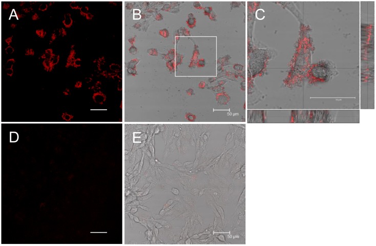
Fig 4. β2-m amyloid fibrils adhere to the cell surfaces.
HIG-82 cells incubated with Ham’s F12 medium containing 100 μg/ml β2-m fibrils (A-C) or vehicle buffer (D, E) for 6 hrs were stained with Congo red and observed with the confocal laser microscope as described in Materials and Methods. (B) and (E) are representative superimposed images on individual bright field micrographs. (C) A higher magnification of the box in (B). Images attached on the right and bottom are those of vertical sections on the lines intersecting at right angles. (A-C) When HIG-82 cells were incubated with 100 μg/ml amyloid fibrils for 6 hrs, they were firmly covered with amyloid fibrils. The scale bars are 50 μm long.

