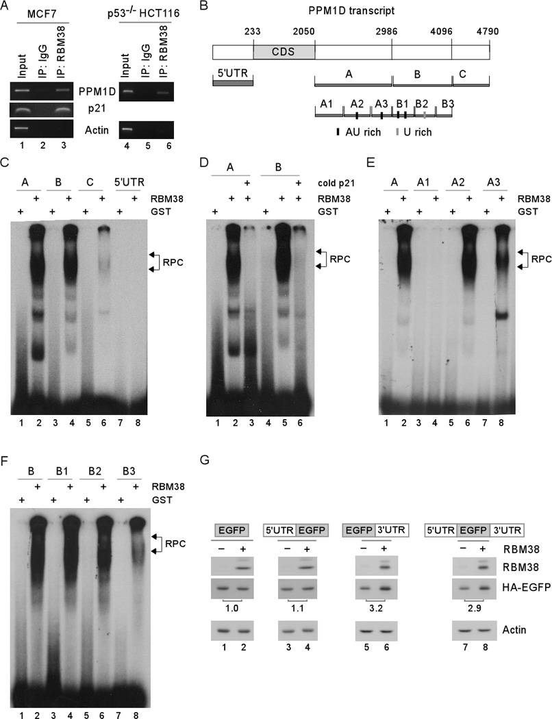
Figure 3. Multiple AU- and U-rich elements in PPM1D 3' UTR are bound by and responsive to RBM38.

(A) RBM38 interacts with PPM1D transcript. Cell extracts purified from MCF7 and p53-null HCT116 cells, which were induced to express RBM38, were immunoprecipitated with a control IgG or RBM38 antibody. The levels of PPM1D and actin transcripts in RBM38 or IgG immunocomplexes were determined by RT-PCR. (B) Schematic presentation of PPM1D transcript and the location of probes used for REMSA. (C) Probes A and B, but not probes C and 5’ UTR, are bound by RBM38. REMSA was performed by mixing 32P-labeled probe A, B, C, or 5’ UTR with recombinant GST or GST-RBM38 protein. The bracket indicates RNA-protein complexes (RPC). (D) Competition assay was performed by adding an excess amount (50-fold) of unlabeled p21 probe derived from p21 3’UTR to the reaction mix prior to incubation with the 32P-labeled probe A or B. (E) REMSA was performed by mixing recombinant GST or GST-RBM38 protein with 32P-labeled probe A, or A1–3. (F) REMSA was performed by mixing recombinant GST or GST-RBM38 protein with 32P-labeled probe B, or B1–3. (G) PPM1D 3′ UTR is responsive to RBM38. An EGFP expression vector, which contains the coding region (ORF) alone or in combination with PPM1D 5′ and/or 3′UTRs, was co-transfected with RBM38-expressing vector into H1299 cells. The levels of EGFP, RBM38, and actin were analyzed by Western blot analysis. The fold change of EGFP was shown below each lane.
