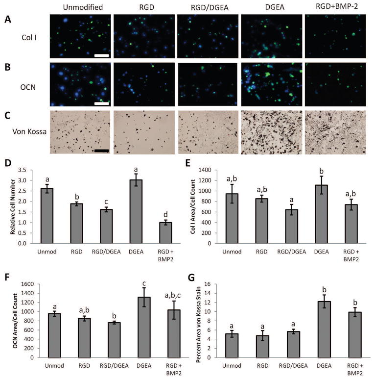
Figure 5. Osteogenic Differentiation of rMSCs Cultured in Peptide-Modified Alginate Hydrogels.
Representative images of rMSCs cultured for 30 days in alginate hydrogels that were either unmodified or modified with RGD and/or DGEA peptides. Recombinant BMP-2, the present clinical standard for inducing bone formation, was included as an additional condition. Cells were stained for (A) collagen I deposition (green), (B) osteocalcin production (green), and (C) mineral deposition (von Kossa). Hoechst 33258 was included as a nuclear counterstain in (B) and (C). Scale bars: 100 μm. (D). Relative number of cells in hydrogels after 30 days in culture as determined by counting Hoechst-stained nuclei. (E). Area of images positive for Col I per cell count. (F). Area of images positive for OCN per cell count. (G). Percent area of images positive for mineralization as indicated by von Kossa staining. In (D–G), conditions with the same letter are not statistically different from each other, p<0.05, Student’s t-test, n = 4–8 independent samples. Error bars are ±SEM.

