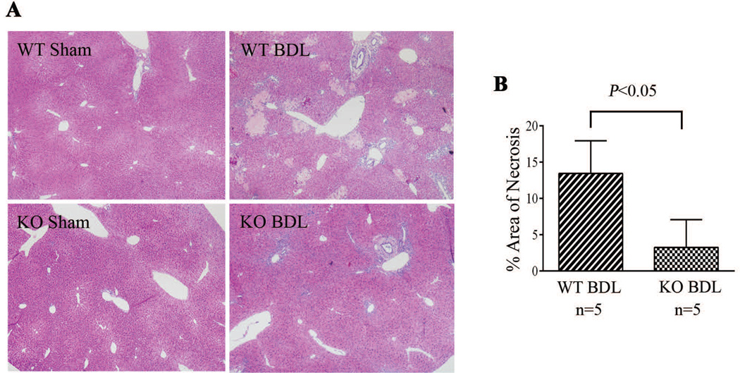
Fig. 5.
Liver necrosis was significantly lower in NHERF-1−/− BDL mice compared with wild-type BDL mice. A. H&E staining of liver sections of sham operated and BDL mice. B. Morphometry of liver necrosis in BDL mice. Data represents means ± SD of 5 animals in each group. Note that no necrosis was detected in both wild-type and NHERF-1−/− sham mouse liver. However, the wild-type BDL group had 14% area of liver necrosis foci, whereas the NHERF-1−/− BDL group had only 3% area of liver necrosis foci. WT: wild-type. KO: NHERF-1−/−.

