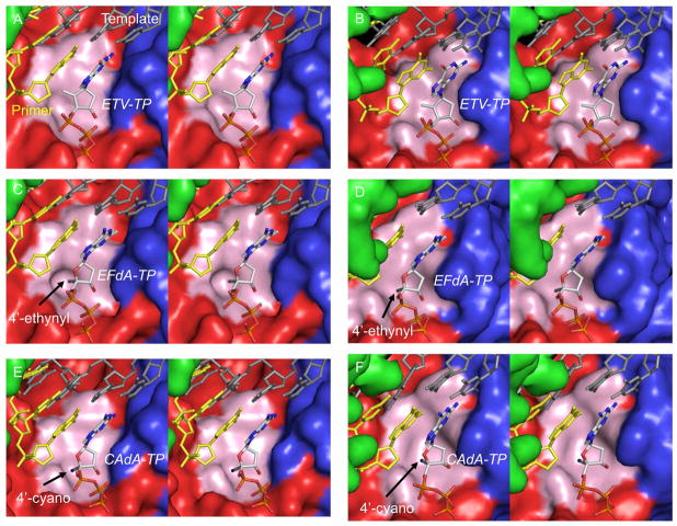
Fig. 5.
Molecular models of HIV-1-reverse transcriptase (RT)/template-primer/NRTI- triphosphates (TP)s and HBV-polymerase (RT)/template-primer/NRTI-TPs. Stereoviews of the ternary complexes of HIV-1-RT/template-primer with ETV-TP (panel A), EFdA-TP (panel C), and CAdA-TP (panel E), are shown in surface representations: fingers, palm, and thumb of RT are colored in blue, red, and green, respectively. Stereoviews of HBV-RT/template-primer with ETV-TP (panel B), EFdA-TP (panel D), and CAdA-TP (panel F) are also shown. The template (dark gray), primer (yellow), and NRTI-TP are rendered as balls-and-sticks. Individual atoms in NRTI-TP are colored as carbon in white, nitrogen blue, oxygen red, phosphorus orange, and fluorine aquamarine. The arrows denote the positions of 4′-ethynyl (panels C and D) and 4′-cyano (panels E and F) moieties. The hydrophobic pocket comprises residues A114, Y115, P157, F160, M184, and D185 in HIV-1-RT and is shown in light pink. In HBV-RT, the pocket is formed by residues A87, F88, P177, L180, M204, and D205 and is shown in light pink. The hydrophobic pocket that accommodates the 4′-susbtituent is deeper in HIV-1-RT than in HBV-RT.

