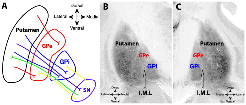
Fig. 1.

A. Schematic of fiber systems traversing the internal (GPi) and external (GPe) segments of the pallidum, including projections to and from the substantia nigra. Labeled pathways include nigrostriatal (green) striatopallidal (red, blue), nigropallidal (yellow), and striatonigral (purple). B and C. Coronal images from the Big Brain atlas, showing the putamen, GPe, and GPi, in the left (B) and right (C) hemispheres; black arrows point to the approximate center of the internal medullary lamina (I.M.L) separating the two segments (slice 3894, Amunts 2013).
