Fig. 2.
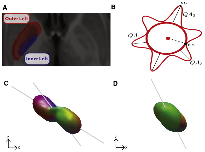
Comparison of the SDFs between the inner and outer semgents of the globus pallidus. A. The inner (blue) and outer (red) segments of the left and right pallidum were manually drawn on the high resolution T1 ICBM 152 template. B. Schematized version of an SDF illustrating three resolved fibers (QA0, QA1, QA2), their magnitude (i.e., lengths, reflecting QA) and orientation. C,D Representative SDFs from the left internal segment (C) and left external segment (D) in the coronal plane from a single subject from the DSI dataset. Gray lines indicate direction of fiber orientations.

