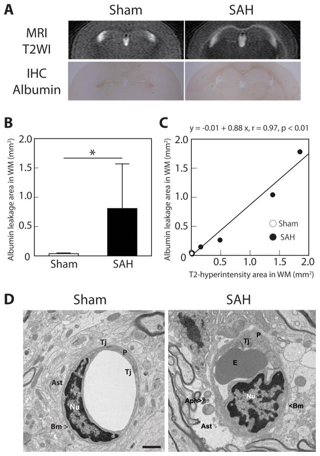
Figure 1.
Subarachnoid hemorrhage (SAH) causes blood-brain barrier disruption at 24 hours after endovascular perforation. (A) Coronal T2-weighted imaging (T2WI) on MRI demonstrated hyperintensity along the white matter (WM) in SAH mice, and this was consistent with albumin leakage demonstrated by immunohistochemistry (IHC). (B) The area of WM albumin leakage was significantly larger after SAH than in sham-operated mice. (C) The area of albumin leakage on IHC correlated with the area of T2-hyperintensity on MRI. Values are mean±SD; *P<0.05; n=4 for each. (D) Transmission electron microscopy demonstrated WM microvascular ultrastructural abnormalities after SAH: swollen astrocyte endfeet with autophagosome, tight junction detachment, erythrocytes trapped in capillary lumen, and irregularity of basement membrane. Aph – autophagosome, Ast – astrocyte, BM – basement membrane, E – erythrocyte, Nu – nucleus of endothelium, P – pericyte, Tj – tight junction. Scale bar = 1μm.

