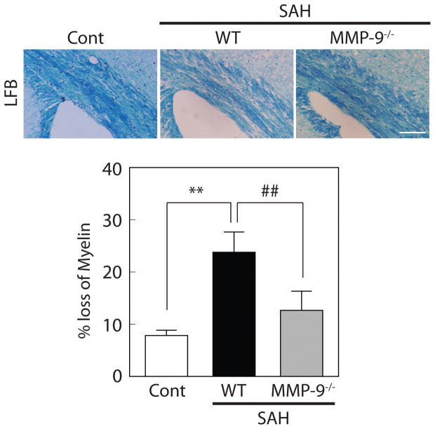
Figure 6.
MMP-9 deletion ameliorated white matter injury after SAH. Representative luxol fast blue (LFB) staining in wild-type (WT) control, WT with SAH, and MMP-9 knockout (MMP-9−/−) mice with SAH at day 8. Values are mean ± SD; **P < 0.01 vs WT control, and ##P < 0.01 vs WT with SAH; n=4 for each. Scale bar = 100μm.

