Fig. 1.

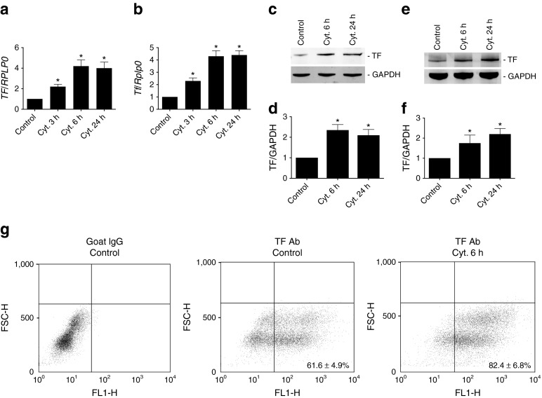
Cytokines induce TF expression in human islets and MIN-6 cells. Human islets and MIN-6 cells were left untreated (Control) or treated with a cytokine mixture of IL-1β + TNF-α + IFN-γ (Cyt.) for the indicated time periods. TF mRNA levels related to RPLP0 in (a) human islets and (b) MIN-6 cells. Immunoblot and graphs showing TF/GAPDH ratio using TF and GAPDH antibodies on (c, d) human islets and (e, f) MIN-6 cells. (g) MIN-6 cells were left untreated (Control) or were treated with cytokine mixture (Cyt.) for 6 h. Cells were incubated with mouse TF antibody (Ab) and TF cell surface expression was analysed using flow cytometry detecting forward scatter height (FSC-H) and fluorescence in fluorescence channel 1 height (FL1-H). Normal goat IgG was used as isotype control. Results are means ± SEM from four (a–f) or three (g) independent experiments. *p < 0.05 vs control, using paired Student’s t test
