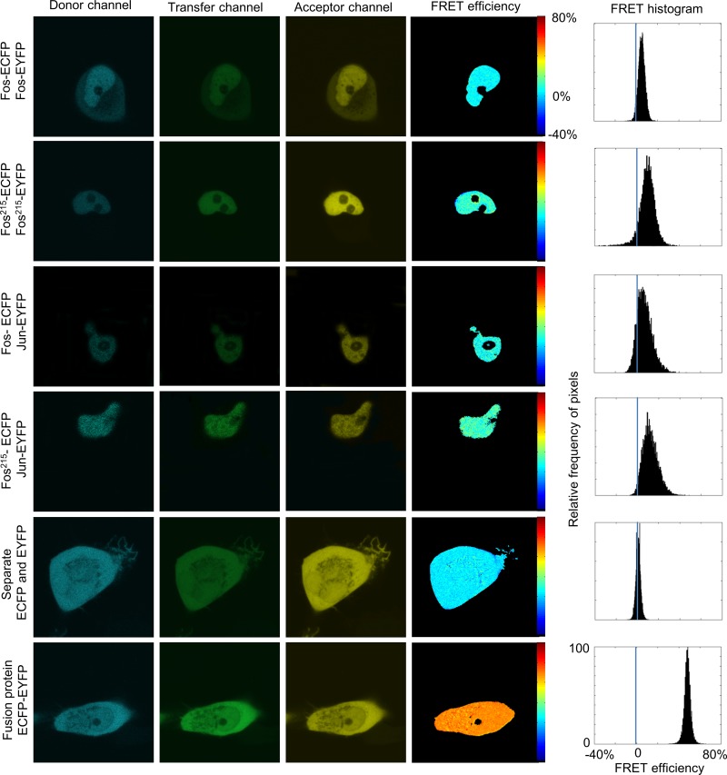
FIG 3.
Subcellular pixel-by-pixel analysis of dimerization by confocal microscopic FRET on HeLa cells. ECFP (donor channel) was excited at 458 nm and detected at 475 to 525 nm. In the transfer channel, excitation was at 458 nm and detection was at 530 to 600 nm. EYFP (acceptor channel) was excited at 514 nm and detected at 530 to 600 nm. Full-length Fos-ECFP–Fos-EYFP (top row), Fos215-ECFP–Fos215-EYFP (second row), Fos-ECFP–Jun-EYFP (third row), and Fos215-ECFP–Jun-EYFP (fourth row) showed nuclear localization. The negative control, ECFP and EYFP expressed independently, and the positive control, the ECFP-EYFP fusion protein (fifth and sixth rows), were evenly distributed in the whole cell. FRET efficiency (E) was calculated in each pixel. Histograms show the statistics of the subcellular distribution of E.

