FIG 4.
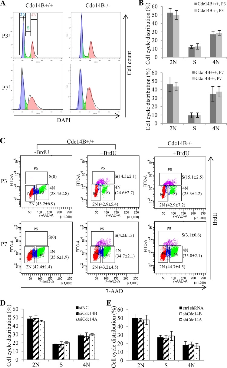
Cdc14 deficiency does not alter cell cycle distribution in the DSB repair assays. (A) Cell cycle analyses of P3 and P7 MEFs by detection of DNA content (DAPI staining) with flow cytometry. (B) Quantification of the cell cycle distribution of the MEFs from panel A. (C) BrdU staining of P3 and P7 MEFs. The MEFs were labeled with 10 μM BrdU for 45 min and stained with FITC-conjugated anti-BrdU. Unlabeled cells were included as negative controls for BrdU staining. Total DNA was stained with 7-AAD for cell cycle analysis by flow cytometry. The numbers (mean ± standard deviation for three independent experiments) indicate the percentage of cells with a DNA content of 2N or 4N and cells in S phase (BrdU positive). (D and E) Cell cycle analyses of the U2OS cells with siRNA (D)- or shRNA (E)-mediated Cdc14 knockdown in the HR assay and NHEJ assay. The error bars indicate standard deviations for at least three independent experiments. ctrl, control. Statistical analyses were performed by unpaired t test (B and C) or one-way ANOVA (D and E). *, P < 0.05; **, P < 0.01.

