FIG 7.
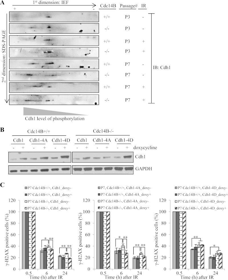
The Cdc14B-regulated Cdh1 phosphorylation level is critical for DSB repair. (A) Cell lysates of P3 and P7 MEFs with or without IR treatment (0.5 h after 10 Gy) were used for 2D gel analyses of Cdh1 phosphorylation levels. + and −, direction of the first-dimension isoelectric focusing. Cdh1 was detected by Western blotting following the second-dimension SDS-PAGE. The spot closer to the “+” end indicates highly phosphorylated Cdh1 with more phosphorylated residues, and the spot closer to the “−” end indicates less phosphorylated Cdh1. (B) Western blotting of induced overexpression of wild-type, phosphodeficient (Cdh1-4A), or phosphomimetic (Cdh1-4D) Cdh1 in MEFs. (C) Quantification of γH2AX-positive cells in late-passage Cdc14B+/+ and Cdc14B−/− MEFs with or without overexpression of wild-type (left panel), phosphodeficient (Cdh1-4A, middle panel), or phosphomimetic (Cdh1-4D, right panel) Cdh1 after 10 Gy of IR. Statistical significance was assessed by ANOVA (*, P < 0.05; **, P < 0.01).

