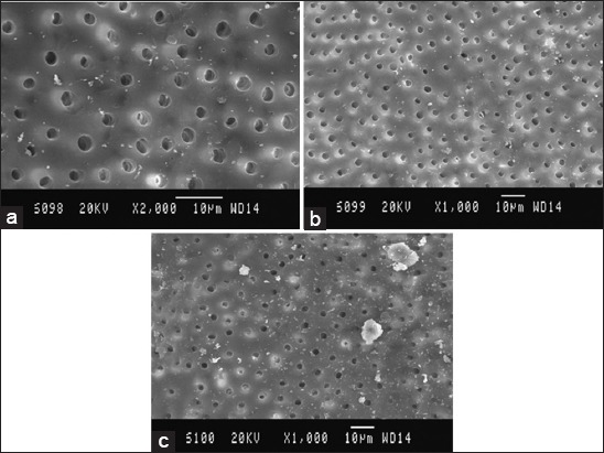
Figure 4.

Scanning electron microscopy (SEM) picture of Group D, (a) Coronal third: SEM picture of the root canal wall of sample in Group D, (b) Middle third: SEM picture of the root canal wall of sample in Group D, (c) Apical third: SEM picture of the root canal wall of sample in Group D.
