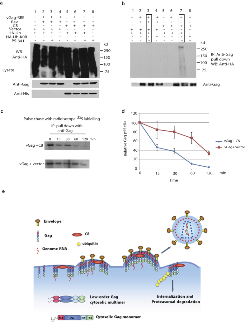
Figure 5. CCDC8-mediated Gag polyubiquitination and degradation.
Western blot analysis of cell lysates (left panel) (a) and co-immunoprecipitation of cell lysate (right panel) (b) with anti-Gag, anti-His and anti-HA. (c) Pulse chase experiment with radioisotope 35S labelled methionine and cysteine. (d) The data in panel (c) were plotted. Error bar represents value ranges from three independent experiments. (e) Model of HIV-1 inhibition by CCDC8.

