

Abstract
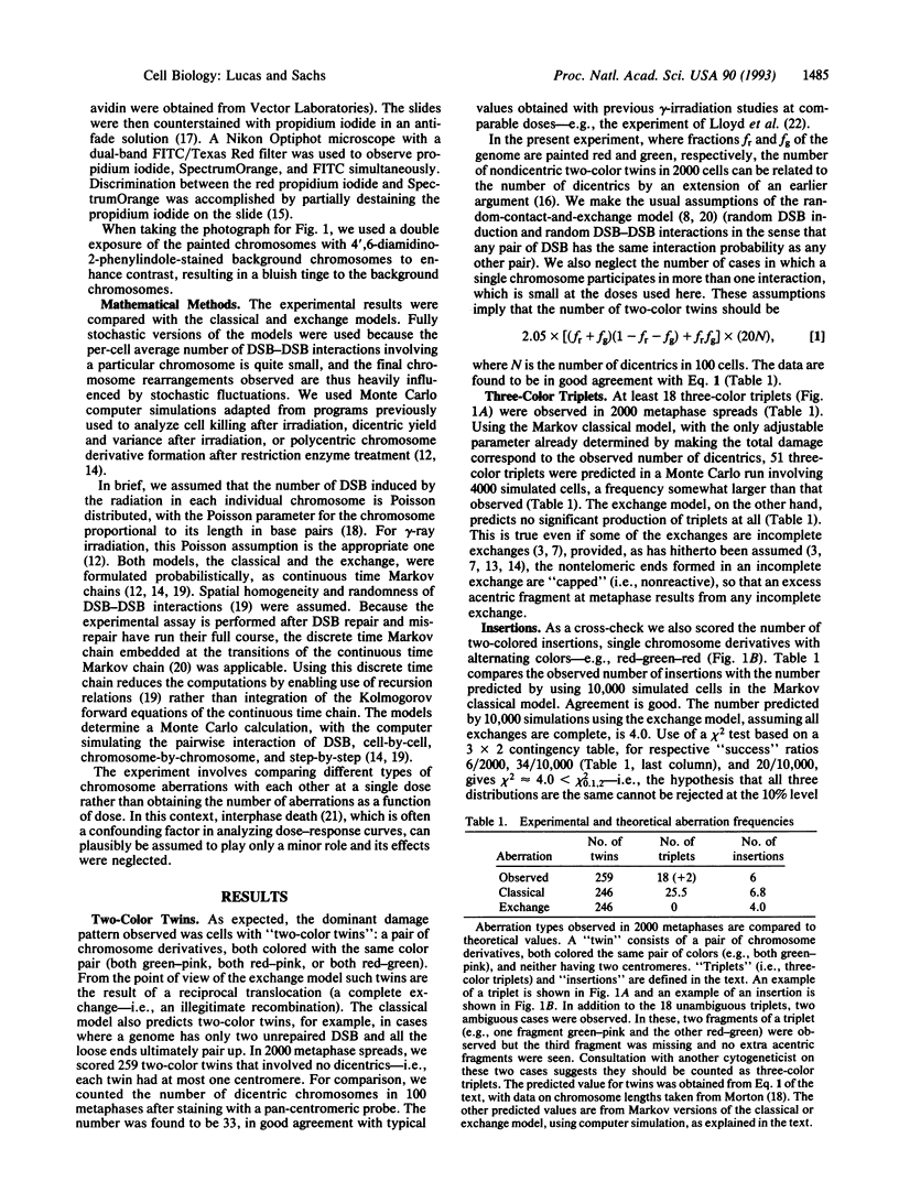
Ionizing radiation induces DNA double-strand breaks (DSB), which interact pairwise to produce chromosome aberrations. There have long been two main competing theories of such pairwise DSB-DSB interactions. The "classical" theory asserts that an unrepaired DSB makes two ends that separate within the cell nucleus, with each end subsequently able to join any similar (nontelomeric) end. The "exchange" theory asserts that at a DSB the chromatin does not separate completely; rather the DSB ends remain associated until repair, or an illegitimate recombination involving another DSB, occurs. The DSB-DSB interaction mechanism was tested by using three-color fluorescence in situ hybridization to paint chromosomes and observe "three-color triplets": three broken and misrejoined chromosomes having cyclically permuted colors. We observed 18 "three-color triplets" in 2000 cells after 2.25 Gy of gamma-irradiation. On the exchange model in its standard form such three-color triplets cannot occur, so this model is inconsistent with the observations. On the classical model, formalized as a discrete time Markov chain embedded at the transitions of a continuous time Markov chain, the frequency of occurrence of three-color triplets can be computed by Monte Carlo simulations. The number of three-color triplets predicted mathematically by the classical model was found to be slightly larger than the observed number. Thus our data, together with our computer simulations, exclude the standard form of the exchange model but are compatible with the classical model. The results are also compatible with other, more complicated models.
Full text
PDF

Images in this article
Selected References
These references are in PubMed. This may not be the complete list of references from this article.
- Bender M. A., Awa A. A., Brooks A. L., Evans H. J., Groer P. G., Littlefield L. G., Pereira C., Preston R. J., Wachholz B. W. Current status of cytogenetic procedures to detect and quantify previous exposures to radiation. Mutat Res. 1988 Sep;196(2):103–159. doi: 10.1016/0165-1110(88)90017-6. [DOI] [PubMed] [Google Scholar]
- Brenner D. J. Track structure, lesion development, and cell survival. Radiat Res. 1990 Oct;124(1 Suppl):S29–S37. [PubMed] [Google Scholar]
- Cornforth M. N., Goodwin E. H. The dose-dependent fragmentation of chromatin in human fibroblasts by 3.5-MeV alpha particles from 238Pu: experimental and theoretical considerations pertaining to single-track effects. Radiat Res. 1991 Jul;127(1):64–74. [PubMed] [Google Scholar]
- Evans J. W., Chang J. A., Giaccia A. J., Pinkel D., Brown J. M. The use of fluorescence in situ hybridisation combined with premature chromosome condensation for the identification of chromosome damage. Br J Cancer. 1991 Apr;63(4):517–521. doi: 10.1038/bjc.1991.123. [DOI] [PMC free article] [PubMed] [Google Scholar]
- Hahnfeldt P., Sachs R. K., Hlatky L. R. Evolution of DNA damage in irradiated cells. J Math Biol. 1992;30(5):493–511. doi: 10.1007/BF00160533. [DOI] [PubMed] [Google Scholar]
- Hlatky L. R., Sachs R. K., Hahnfeldt P. The ratio of dicentrics to centric rings produced in human lymphocytes by acute low-LET radiation. Radiat Res. 1992 Mar;129(3):304–308. [PubMed] [Google Scholar]
- Hlatky L., Sachs R., Hahnfeldt P. Reaction kinetics for the development of radiation-induced chromosome aberrations. Int J Radiat Biol. 1991 May;59(5):1147–1172. doi: 10.1080/09553009114551041. [DOI] [PubMed] [Google Scholar]
- Johnson G. D., Nogueira Araujo G. M. A simple method of reducing the fading of immunofluorescence during microscopy. J Immunol Methods. 1981;43(3):349–350. doi: 10.1016/0022-1759(81)90183-6. [DOI] [PubMed] [Google Scholar]
- Lucas J. N., Awa A., Straume T., Poggensee M., Kodama Y., Nakano M., Ohtaki K., Weier H. U., Pinkel D., Gray J. Rapid translocation frequency analysis in humans decades after exposure to ionizing radiation. Int J Radiat Biol. 1992 Jul;62(1):53–63. doi: 10.1080/09553009214551821. [DOI] [PubMed] [Google Scholar]
- Lucas J. N., Poggensee M., Straume T. Translocations between two specific human chromosomes detected by three-color "chromosome painting". Cytogenet Cell Genet. 1993;62(1):11–12. doi: 10.1159/000133434. [DOI] [PubMed] [Google Scholar]
- Morton N. E. Parameters of the human genome. Proc Natl Acad Sci U S A. 1991 Sep 1;88(17):7474–7476. doi: 10.1073/pnas.88.17.7474. [DOI] [PMC free article] [PubMed] [Google Scholar]
- Prosser J. S., Edwards A. A., Lloyd D. C. The relationship between colony-forming ability and chromosomal aberrations induced in human T-lymphocytes after gamma-irradiation. Int J Radiat Biol. 1990 Aug;58(2):293–301. doi: 10.1080/09553009014551631. [DOI] [PubMed] [Google Scholar]
- Sachs R. K., Yates B. L., Tarver J., Morgan W. F. Modelling the formation of polycentric chromosome aberrations. Int J Radiat Biol. 1992 Oct;62(4):449–460. doi: 10.1080/09553009214552331. [DOI] [PubMed] [Google Scholar]
- Savage J. R., Harvey A. N. Revell revisited. Mutat Res. 1991 Sep-Oct;250(1-2):307–317. doi: 10.1016/0027-5107(91)90186-r. [DOI] [PubMed] [Google Scholar]
- Savage J. R. The production of chromosome structural changes by radiation: an update of Lea (1946), Chapter VI. Br J Radiol. 1989 Jun;62(738):507–520. doi: 10.1259/0007-1285-62-738-507. [DOI] [PubMed] [Google Scholar]
- Sax K. An Analysis of X-Ray Induced Chromosomal Aberrations in Tradescantia. Genetics. 1940 Jan;25(1):41–68. doi: 10.1093/genetics/25.1.41. [DOI] [PMC free article] [PubMed] [Google Scholar]