Figure 2.

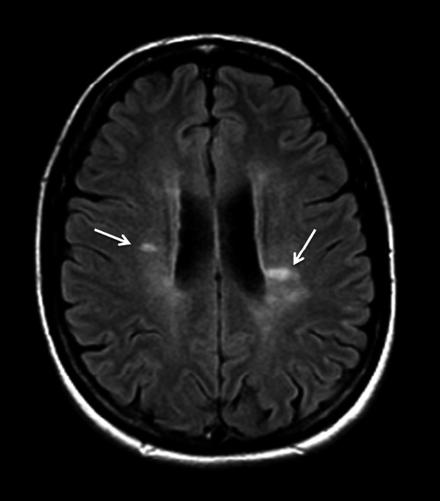
Brain MRI axial fluid attenuated inversion recovery (FLAIR) image shows the characteristic periventricular areas of increased signal intensity (arrows) that are oriented perpendicular to and often contiguous with the lateral ventricles.

Brain MRI axial fluid attenuated inversion recovery (FLAIR) image shows the characteristic periventricular areas of increased signal intensity (arrows) that are oriented perpendicular to and often contiguous with the lateral ventricles.