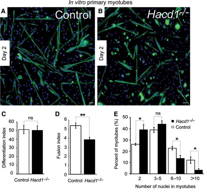
Figure 4.
Defective myoblast fusion in normally differentiating HACD1-deficient myoblasts. (A and B) Immunodetection on muscle primary cell cultures from control or Hacd1−/− newborns on Day 2 of differentiation of the myosin heavy chains (MHC, green). Nuclei are in blue. Scale bar, 100 μm. (C) Unchanged differentiation index in HACD1-deficient myoblasts, i.e. percentage of nuclei contained in MHC-positive cells (n = 5 for each newborn group; ≥1000 nuclei per sample). (D) Decreased fusion index in HACD1-deficient myoblasts, i.e. average number of nuclei per myotube (n = 5 for each newborn group; ≥100 myotubes per sample). (E) Percent distribution of myotubes analysed in D by their number of nuclei. Error bars correspond to standard error of the mean. *P < 0.05, **P < 0.01, ***P < 0.001.

