Fig. 6.

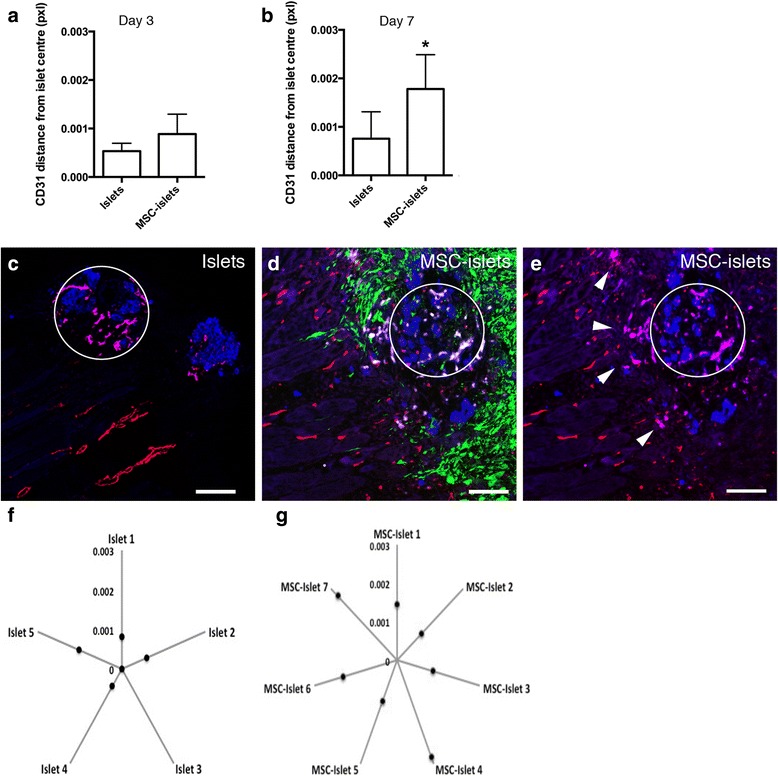
Facilitated islet endothelial cell migration out into the surrounding tissue in the presence of MSC. a Mean distance of human CD31 expressing vessels from the center of the islet in control islets and MSC-islets at three days post transplantation (n = 4). b Mean distance of human CD31 expressing vessels from the center of the islet in control islets compared to MSC-islets at seven days post transplantation (*p < 0.05, islets n = 5, MSC-islets n = 7) using a Mann–Whitney test. c In control sections after one week, the human islet endothelial cells (purple) remained inside the islet graft (blue, centrally marked by a white circle) avoiding direct contact with the recipient vasculature (red). d In the MSC-islets (green and blue, respectively, centrally marked by a white circle) the presence of human islet endothelial cells (white/purple) were more prominent at distance of the islet graft at the same time point. e Same figure as in panel D excluding the MSC signal in green showing the human islet endothelial cells (purple, marked with white arrow heads) migrating out to the surrounding tissue (day 7). f Mean distance of human endothelial cell migration from the center of the control islet grafts in each study object 7 days post transplantation (n = 5). g Mean distance of human endothelial cell migration from the center of the MSC-islet grafts in each study object 7 days post transplantation (n = 7). Bars = 100 um
