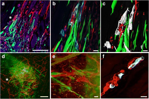
Fig. 7.

Cellular interactions between MSC and human islet endothelial cells with the recipient vasculature one week post transplantation. a Interactions between MSC (green) and the CD31 expressing human islet endothelial cells (light blue) with the recipient vasculature (red). b Increased magnification in asterisk marked area of panel A showing the interactions between the MSC and human islet endothelial cells where the MSC are wrapped around the mouse vasculature. c Imaris visualization of the image shown in panel b. d In situ staining and in situ confocal microscopy created an overview of the graft area showing sprouting MSC (green) close to the mouse vasculature (red). e Increased magnification of the asterisk area in panel d showed MSC in green wrapped around the mouse vasculature in red. f Formation of a chimeric blood vessel in the close proximity of the MSC. The human islet endothelial cells (white) are fused with the murine blood vessel (red). Bars = 100 um in panel a and d, 5 um in panel b and c and 10 um in panel e and f
