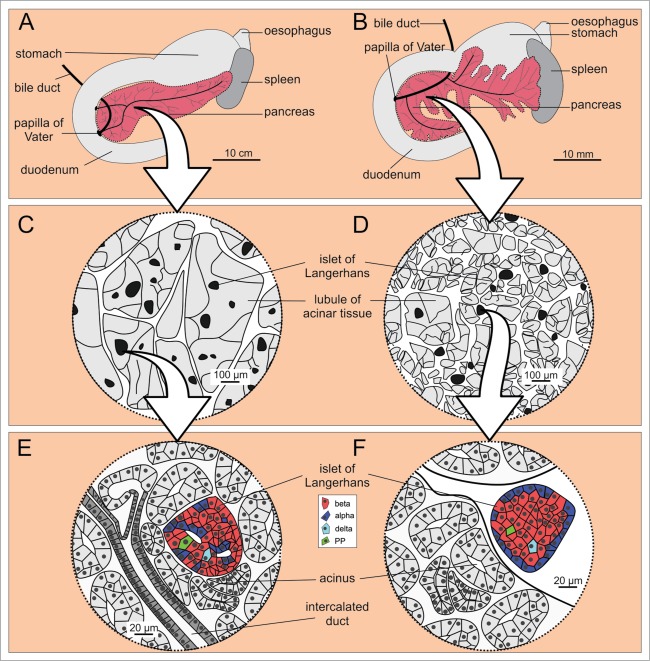
Figure 2.
Microscopic anatomy of the human (A, C, and E) and the mouse (B, D, and F) pancreas. (A and B) Macroscopic anatomy of the human and the mouse pancreas, respectively. (C and D) Magnifying a portion of pancreas reveals larger lobules in humans when compared to mouse, whereas the islets of Langerhans are of fairly comparable size in humans and mice. (E and F) Cell composition and location of the islets of Langerhans within the pancreas are markedly different in the 2 species. Note the more diffusely distributed endocrine cells in humans (E) and the mantle-core pattern in mice (F).

