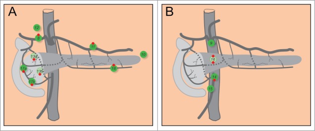
Figure 5.

Lymph nodes of the pancreas. (A) Groups of lymph nodes that outline the gland. (B) Groups of nodes at the aorta. 8: hepatic nodes. 9: celiac nodes. 10: splenic and gastrosplenic nodes. 11: suprapancreatic nodes. 12: hepatoduodenal nodes. 13a: superior posterior nodes. 13b: inferior posterior nodes. 14: superior mesenteric nodes. 15: middle colic nodes. 16: paraaortic nodes. 17a: superior anterior nodes. 17b: inferior anterior nodes. 18: infrapancreatic nodes. Red dots on the top and at the bottom of lymph nodes indicate the nodes most frequently involved in carcinoma of the body or the tail, and the head, respectively.
