
Figure 6.
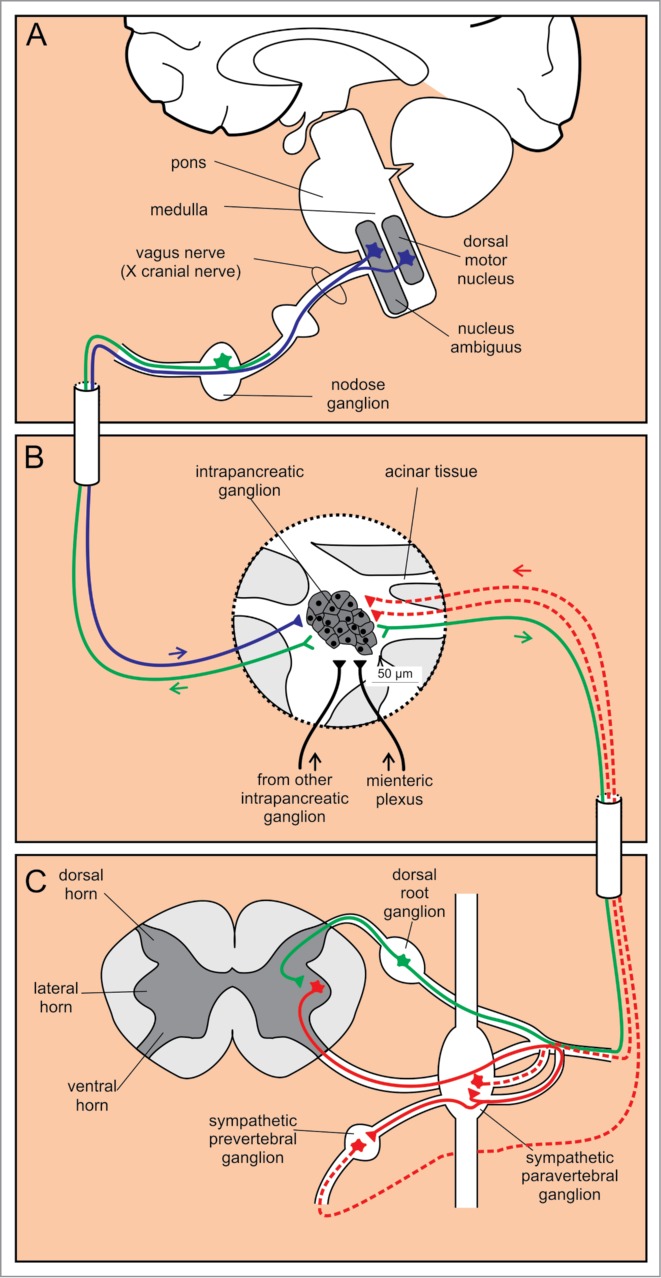
Autonomic innervation of intrapancreatic ganglia. (A) Cell bodies of efferent parasympathetic fibers are located in 2 nuclei in the medulla, whereas the afferent sensory cell bodies are positioned in the nodose ganglion. (B) Intrapancreatic ganglia receive input from sympathetic (red) and parasympathetic (blue) fibers, from other intrapancreatic ganglia and from the myenteric plexus (black). Additionally, sensory fibers (green) project from intrapancreatic ganglia. (C) Preganglionic efferent sympathetic fibers (red solid line) project from cell bodies in the lateral horn of the spinal cord to paravertebral and prevertebral sympathetic ganglia. From here, the postganglionic sympathetic fibers (red dashed lines) project to the intrapancreatic ganglia. Sensory afferent fibers (green) have their cell bodies in the dorsal root ganglia.