
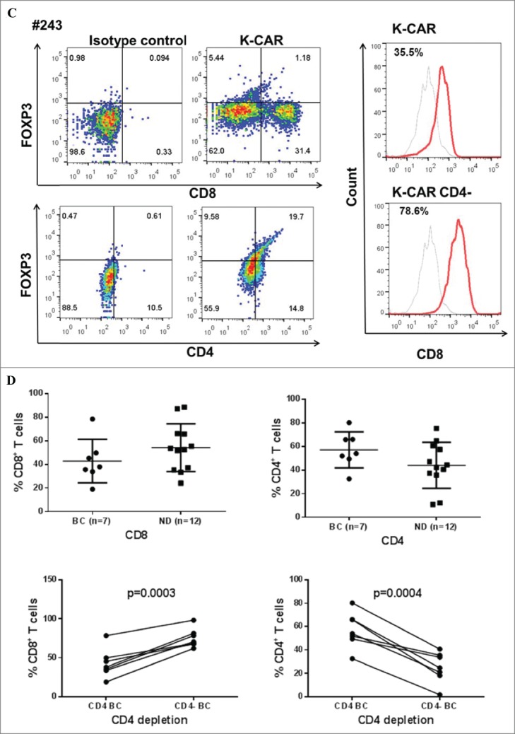
Figure 1 .
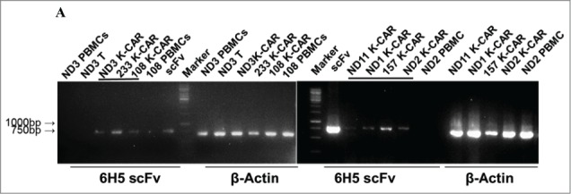
Characterization of K-CAR in various donors. (A) RT-PCR was employed to detect the expression of 6H5 scFv (700 bp) using scFv specific primers. Amplified β-actin was used as a loading control, and scFv plasmid was used as a positive control. Expression of 6H5 scFv was demonstrated in K-CAR T cells obtained from BC patients and NDs. No scFv expression was detected in control T cells or PBMCs. (B) Both Fc+ and CD3+ T cell populations were determined in T cells transfected with K-CAR or GFP from patient 157 (top panel) and ND1 (bottom panel) by FACS using anti-Fc and CD3 antibodies on days 7, 21 and 35 post-transfection. The isotype alone was used as control. (C) Both FOXP3+ and CD8+ or CD4+ T cells from BC patient 243 were determined by FACS (left panel). The percentage of CD8+ T cells was increased in K-CAR T cells after CD4+ depletion (right panel). (D) Lower percentages of CD8+ and higher percentages of CD4+ T cells were demonstrated in K-CAR T cells obtained from BC patients than from NDs (top panel). Significantly enhanced CD8+ (P = 0.0003) and reduced CD4+ (P = 0.0004) T cell populations were demonstrated in T cells from BC patients (n = 7) after CD4 depletion.
Figure 1.

(Continued)
Figure 1.

(Continued)
Figure 1.
(Continued)