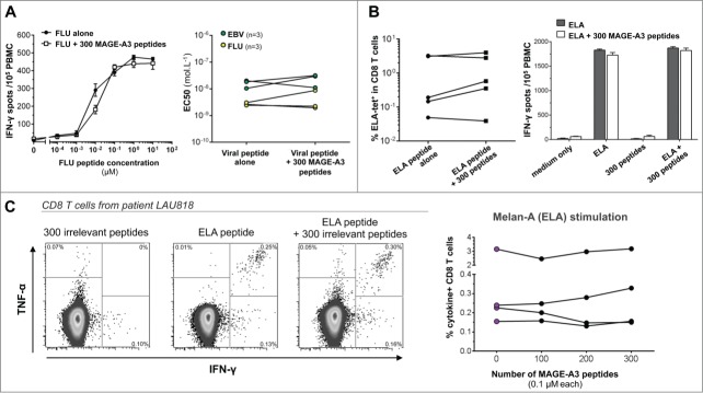
Figure 2.

The presence of 300 irrelevant peptides does not influence the detection of viral and tumor specific T-cell responses. (A) Ex vivo TCR avidity of HLA-A2 restricted FLU58–66 and CMV495−503 specific CD8+ T-cell responses. The left panel shows representative data of the responsiveness of anti-FLU T cells after stimulation with limiting peptide dilutions (10 to 10−4 μM), in the presence or absence of 300 MAGE-A3 peptides. The right panel depicts cumulative data from six donors correlating the EC50 values of PBMC stimulated with the specific peptide alone or mixed with a pool of 300 MAGE-A3 peptides. (B) In vitro Melan-A26–35-specific T-cell proliferation and tetramer staining. PBMC from healthy donors were cultured for one week in the presence of either ELA peptide alone or ELA plus the MAGE-A3 peptide pool. At day 7, cells were collected and stained with the Melan-A26–35 tetramer (left panel) or boosted in different conditions to check for their specificity by IFNγ-ELISpot assay (right panel). Black bars show mean ± SEM response of cells stimulated for one week with ELA alone, white bars show mean ± SEM response of cells stimulated with ELA together with the pool. (C) Sensitive ex vivo detection of Melan-A26–35-specific T cells in vaccinated melanoma tumor patients. PBMCs were stimulated overnight with increasing numbers of MAGE-A3 peptides (up to 300) and then tested for cytokine production. The left panels show representative ICS analysis of PBMCs from one out of four patients tested. The right graph depicts cumulative data showing the percentages of cytokine-producing CD8+ T cells in the indicated conditions.
