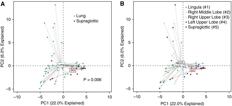
Figure 3.
Ordination of supraglottic and intrapulmonary bacterial communities. (A and B) Unsupervised ordination (principal components analysis) of supraglottic (SG) versus combined intrapulmonary specimens (A) or individual intrapulmonary specimens (B), labeled by specimen site. Numbers in B refer to the order of sampling during bronchoscopy. Supraglottic communities were significantly distinct from lungs collectively (P = 0.006) and, with the exception of the right upper lobe (RUL), individually (see Table 2 for significance). LUL = left upper lobe; RML = right middle lobe.

