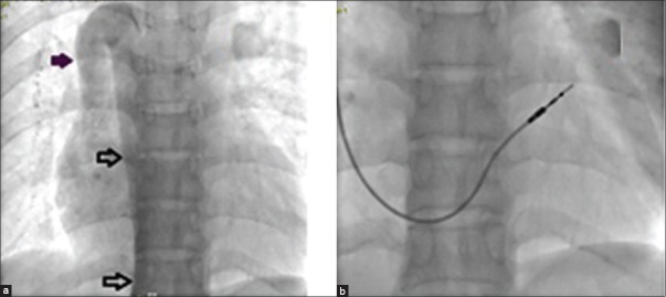
Figure 4.

(a) Venogram through right femoral venous access shows Interrupted inferior vena cava with azygos continuation draining into superior vena cava (bold arrow) (b) Permanent pacemaker implanted with a right ventricle lead positioned at outflow tract
