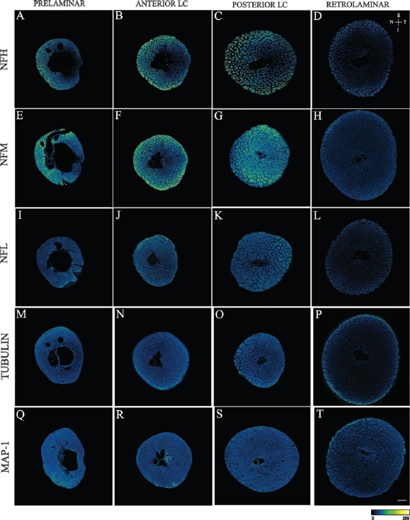
Figure 3.

Confocal microscope images of the prelaminar, anterior lamina cribrosa (anterior LC), posterior lamina cribrosa (posterior LC) and retrolaminar regions of human optic nerves stained with antibodies to neurofilament heavy subunit (NFH) (A to D), neurofilament medium subunit (NFM) (E to H), neurofilament light subunit (NFL) (I to L), Tubulin (M to P) and microtubule associated protein (MAP)-1 (Q to T).
Images are pseudo coloured according to the pixel intensity scale presented at the bottom of the images. Arrows allow orientation of the superior (S), inferior (I), nasal (N) and temporal (T) margins of the images. Scale bar: 300 μm. Note: Reproduced from Kang et al., 2014, Sectoral variations in the distribution of axonal cytoskeleton proteins in the human optic nerve head. Exp Eye Res 128:141-150.
