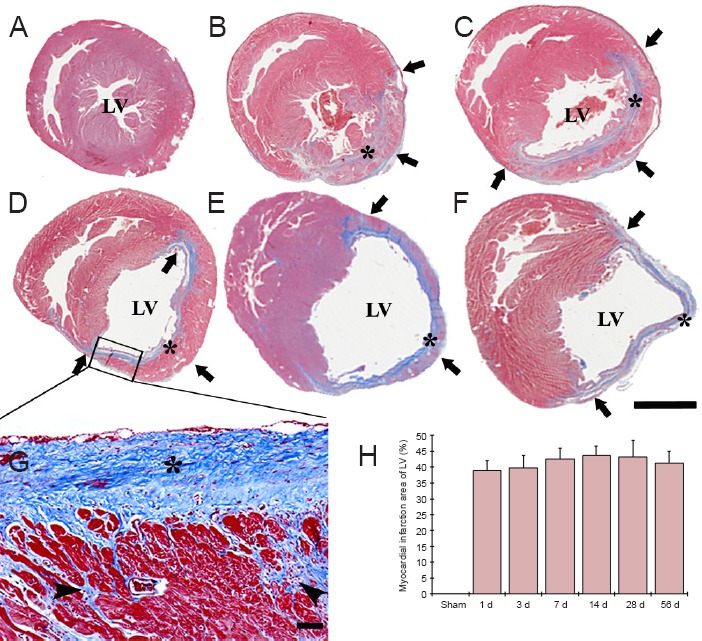
Figure 1.

Masson's trichrome staining of the left ventricle (LV) wall in the sham-operated (sham) rats (A) and myocardial infarction (MI) rats at 3 (B), 7 (C), 14 (D, G), 28 (E), 56 days (F) after coronary artery ligation.
Ventricular dilation was observed in the LV after MI, the infarcted wall of the LV became thin (arrows) after MI, while the non-infarcted wall showed hypertrophy. Collagen (asterisks) was observed in the infarcted wall from 3 days after MI, and the collagen accumulated with time after MI (G). Collagen (asterisks) and myofibroblasts (arrowheads) continually accumulated after MI, and replaced by necrotic myocardium after MI (G). Scale bars: 5 mm in A–F, 50 μm in G. (H) The area of myocardial infarction in the LV (n = 7 rats per group). There was no significant difference in the area of myocardial infarction between each time point following MI. The bars indicate the mean ± SEM. d: Day(s).
