Fig. 1.
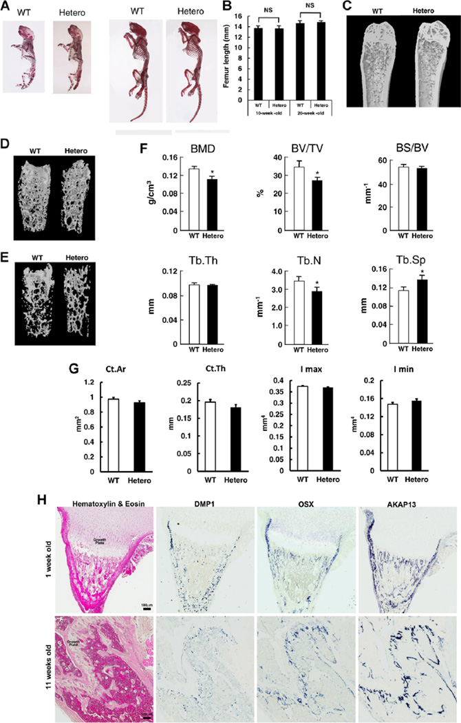
Bone phenotype of Akap13+/− mice and expression of Akap13 in bone. (A) Skeletal structure of WT and Akap13+/− mice (Hetero). Left: newborn mice. Right: 10-week-old mice. Skeletal staining by alizarin red. (B) Bone length in 10-week-old and 20-week-old Akap13+/− mice compared with WT mice. Bars represent the mean ± SE (n = 5). (C) Femurs from 20-week-old WT and Akap13+/− mice revealed decreased bone mineral density. (D, E) 3D rendering images of µCT of femora from 20-week-old WT or Akap13+/− mice showed decreased trabecular bone in the Akap13+/− mice. (F) Quantitative comparison of femoral microstructure parameters of 20-week-old WT and Akap13+/− mice. Global threshold method was used with a fixed range of grayscale values for solid pixels (white) and pixels outside this range were set as space pixels (black). Voxels range from 0 (air; HV-1000) to 255 (HV16444) with water 14.618 on the grayscale voxel. The threshold was a lower of 24 and upper of 255 voxels, the number of layers was 101, which was used for all specimens. *p < 0.05 (n = 5 mice/group). (G) Cortical parameters measured by µCT for the distal femur of 20-week-old female WT and Akap13+/− mice. (H) In situ hybridization for Akap13 transcripts. Formalin fixed, decalcified sections from proximal tibias of 1-week-old (upper panels) or 11-week-old (lower panels) mice were hybridized to DIG-labeled riboprobes for Akap13, Osterix (Osx), or Dentin matrix phosphoprotein 1 (Dmp1), producing a purple color visualized with a ScanScope CS digital scanner under bright field microscopy. Left: Hematoxylin and eosin–stained sections. Right: In situ hybridization (not counterstained). Akap13 localization in the tibia resembled that of Osterix, a specific marker for preosteoblasts and osteoblasts. BMD = trabecular bone mineral density; BV/TV = trabecular bone volume per tissue volume; BS/BV = bone surface per bone volume; Tb.Th = trabecular thickness; Tb.N = trabecular number; Tb.S = trabecular spacing; Ct.Ar = cortical area; Ct.Th = cortical thickness; Hetero = Akap13+/− mice.

