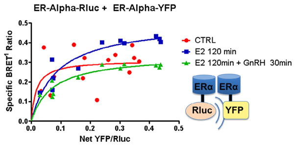
Fig. 3.

Effect of GnRH on ERα dimerization. ERα-Rluc + ERα-YFP: GT1-7 cells in serum-and phenol red-free 1:1 DMEM/F12 medium were transiently transfected with 0.2 μg of ERα-Rluc and increasing amounts from 0 to 1.0 μg of ERα-YFP (y-axis). BRET1 measurements were then taken in triplicate of untreated cells (x-axis), cells treated with E2 100 nM for 2 hours or cells treated with E2 100 nM for a total of 120 min, with GnRH agonist 100 nM added for the last 30 min of incubation. Saturation curves were then plotted as shown, demonstrating a decrease in ligand-induced ERα–ERα dimerization (dotted line, square boxes) when cells were also treated with GnRH (BRETmax 0.311 ± 0.046 vs. 0.507 ± 0.045 vs 0.336 ± 0.025, p = 0.018).
