
Figure 1.
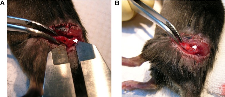
An animal model of bone defects.
Notes: Male C57LB/L mice were anesthetized, and a metaphyseal bone defect was drilled in the proximal femur (A). After rinsing in phosphate-buffered saline and subsequent exposure to ultraviolet light for 30 minutes, chitosan nanofiber scaffolds were implanted into the bone defect (B). The wound was sutured, and the animals were allowed free unrestricted weight-bearing after recovery from anesthesia. Only one defect was created in each proximal femur of an animal, and totally nine animals were treated in this study. Arrows indicate the bone defect sites.