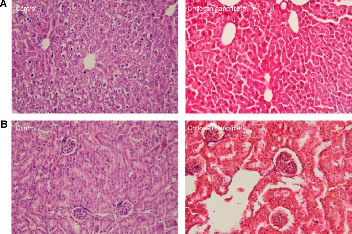
Figure 3.
Toxicities of chitosan nanofibers to the liver and kidneys.
Notes: Bone defects were surgically created in the proximal femurs of male C57LB/L mice, and chitosan nanofibers were implanted into one defect for 21 days. After that period, animals were sacrificed, and the liver and kidneys were removed, cleaned, and weighed. These samples were fixed with paraformaldehyde and embedded in paraffin. Following slicing, liver (A) and kidney (B) specimens prepared from control (left panels) and chitosan nanofiber-treated (right panels) animals were stained with hematoxylin and eosin and observed and photographed under a light microscope at 200×. Only one defect was created in each proximal femur of an animal, and totally nine animals were treated in this study.

