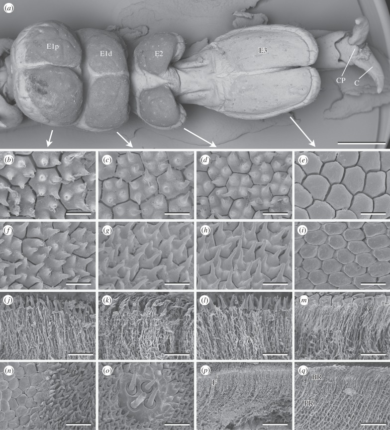
Figure 1.
Tarsal structures of A. diadematus. (a) Ventral view of the tarsus. Surface structures (b–i,n,o) and fractures (j–m,p,q) of the proximal portion of the first euplantula (b,f,j,n,o), distal portion of the first euplantula (c,g,k), second (d,h,l,p), and third (e,i,m,q) euplantula. (n) Transition from the outer zone to the nubby structures. (o) Aggregate of sensilla. E1p, E1d, proximal and distal euplantula of tarsomere 1; E2 and E3, euplantulae of tarsomeres 2 and 3; C, claw; CP, claw pad; BR, layer of branching rods; F, layer of fine filaments; PR, layer of principal rods. Scale bars: (a) 1 mm, (b–i) 5 µm, (j–m) 10 µm, (n,o) 15 µm and (p,q) 30 µm.

