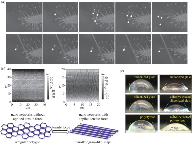
Figure 4.
Characterization of the sundew adhesive. (a) Images captured by the high-speed camera system (Powerview HS-650), demonstrating the viscoelastic adhesion of sundew adhesive. Microscope coverslips were in situ in contact with the adhesive droplets on the leaves of sundew plants. Arrows indicate the flat discs formed by sundew adhesive upon touching the substrates. (b) Upper panel, AFM images of the nano-network architectures in sundew adhesive before (left) and after (right) a tensile force was applied. Image sizes are 40 × 40 µm (left) and 20 × 20 µm (right), with height bars. Bottom panel, a schematic drawing of the deformation of the nano-network structures within sundew adhesive, in response to the exerted external force. (c) CA measurements. Upper panel, water and sundew adhesive were dropped onto respective siliconized glass slides. Middle panel, water was dropped onto bare siliconized glass slides and sundew adhesive-coated siliconized glass slides, respectively. Bottom panel, water was dropped onto bare polystyrene slides and sundew adhesive-coated polystyrene slides, respectively. (Online version in colour.)

