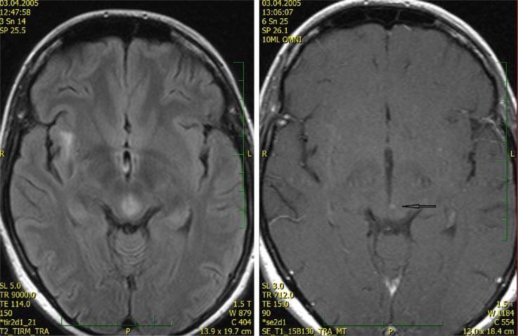
Figure 4.
MRI at time of neurological onset 2 (2005).
Notes: The infiltrative lesion is situated around the cerebral aqueduct in the mesencephalon. Only very discrete dot-like enhancement is apparent after gadolinium administration (arrow). (Native FLAIR and post-gadolinium SE T1 WI, transversal).
Abbreviations: MRI, magnetic resonance imaging; FLAIR, fluid attenuation inversion recovery; SE T1 WI, spin echo T1 weighted images.

