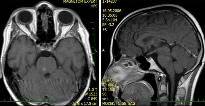
Figure 6.
MRI at time of neurological progression 2 (2006).
Notes: Small ring-like post-gadolinium enhancement in the brain stem lesion also depicted in Figure 5. (Post-gadolinium SE T1 WI, transversal and sagittal).
Abbreviations: MRI, magnetic resonance imaging; SE T1 WI, spin echo T1 weighted images.

