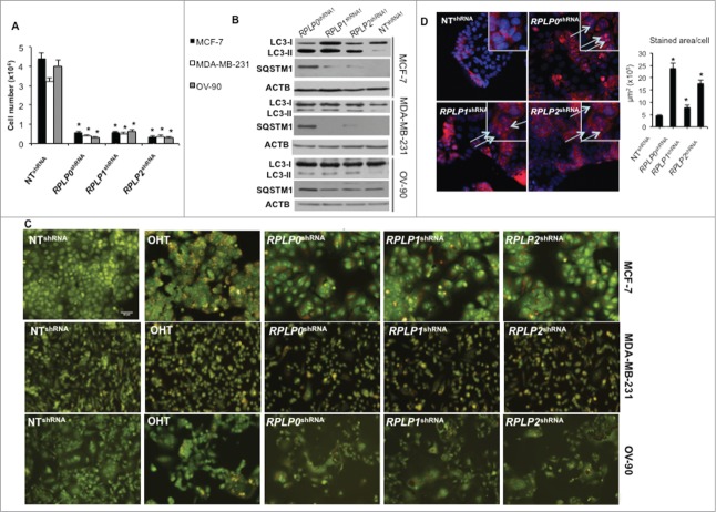
Figure 2.
RPLP protein downregulation induces autophagy in breast and ovarian cancer cell lines. (A) Numbers of MCF-7, MDA-MB-231, and OV-90 cells after transfection with RPLP0 shRNA, RPLP1 shRNA, RPLP2 shRNA, or NT shRNA vector at the 3rd d of selection with puromycin. *, P ≤ 0.01. The data are the mean ±SD of 3 independent experiments. (B) Representative immunoblot for autophagy, which is characterized by the conversion of LC3-I (cytosolic form) into LC3-II (autophagosome membrane-bound form), and SQSTM1. Note the decrease of SQSTM1 protein and the increase in LC3-II levels compared with ACTB in MCF-7, MDA-MB-231, and OV-90 cells expressing RPLP0 shRNA, RPLP1 shRNA, RPLP2 shRNA, or NT shRNA vector at 72 h after transfection. (C) Acridine orange staining of MCF-7, MDA-MB-231, and OV-90 cells stably expressing shRNAs (as in A). Cells treated with 2.5 µM OHT for 4 d were used as a positive control for acidic vesicles, in particular the autolysosomes characteristic of autophagy. (D) MCF-7 cells expressing RPLP0 shRNA, RPLP1 shRNA, RPLP2 shRNA, or NT shRNA vector were incubated with BECN1 antibody and then analyzed using a fluorescence microscope. Arrows signal the punctate staining indicating BECN1 expression. Positive staining was scored in 100 cells, with error bars indicating mean values ±SD. *, P ≤ 0.05.

