Figure 5.
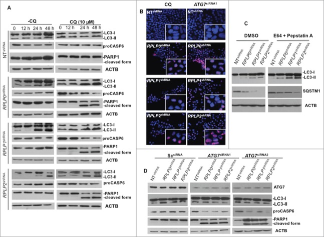
Autophagy inhibition triggers apoptotic cell death. (A) MCF-7 cells were treated at different time points with CQ, an autophagy inhibitor that prevents the fusion of the autophagosome with the lysosome to form the autolysosome, a late stage in the autophagy cascade. At 24 h and 48 h, CQ was able to prevent autophagy in MCF-7 cells depleted of RPLP0, RPLP1, or RPLP2 while stimulating apoptosis, as observed by the decrease in proCASP6 and the presence of the cleaved PARP1 form characteristic of apoptosis. (B) Immunofluorescence of PARP1 staining in MCF-7 cells treated with ATG7 siRNA with deletion of the indicated RPLP protein. Staining in control cells (NT shRNA) was concentrated in the nucleus (acting as a reservoir). In MCF-7 cells depleted of each RPLP protein (RPLP0, RPLP1, or RPLP2), PARP1 staining was diffuse throughout the nucleus. The morphological appearance of cells treated with CQ is also shown. (C) Addition of E64/pepstatin A in cells depleted of RPLP0, RPLP1 or RPLP2 proteins results in a greater increase of LC3-II and diminishes the degradation of SQSTM1. (D) Effect of 2 ATG7 siRNAs on LC3-II conversion, proCASP6, and the cleaved PARP1 form in MCF-7 cells depleted for the indicated RPLP protein vs. control cells (NT shRNA) and Sc siRNA. These results indicate that ATG7 siRNA is able to avoid the autophagic phenotype by stimulating apoptosis.

