Figure 7.
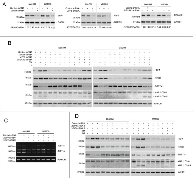
XBP1 plays an important role in RIPK1 upregulation in melanoma cells upon ER stress. (A) Whole cell lysates from Mel-RM and MM200 cells transduced with the control, ERN1, ATF6, or EIF2AK3 shRNA were subjected to western blot analysis of ERN1 (left panel), ATF6 (middle panel) or EIF2AK3 (right panel) and GAPDH (as a loading control). The numbers represent knockdown efficiencies. The data shown are representative of 3 individual western blot analyses. (B) Mel-RM and MM200 cells transduced with the control, ERN1, ATF6, or EIF2AK3 shRNA were treated with tunicamcyin (TM) (3 μM) or thapsigargin (TG) (1 μM) for 16 h. Whole cell lysates were subjected to western blot analysis of HSF1, RIPK1, SQSTM1, MAP1LC3A, and GAPDH (as a loading control). The data shown are representative of 3 individual experiments. (C) Mel-RM and MM200 cells transfected with the control or XBP1 siRNA were subjected to RT-PCR for the analysis of XBP1 mRNA. GAPDH was used as a loading control. The data shown are representative of 3 individual qPCR analyses. (D) Mel-RM and MM200 cells transfected with the control or XBP1 siRNA were treated with TM (3 μM) or TG (1 μM) for 16 h. Whole cell lysates were subjected to western blot analysis of HSF1, RIPK1, SQSTM1, MAP1LC3A, and GAPDH (as a loading control). The data shown are representative of 3 individual experiments.

