Figure 3.

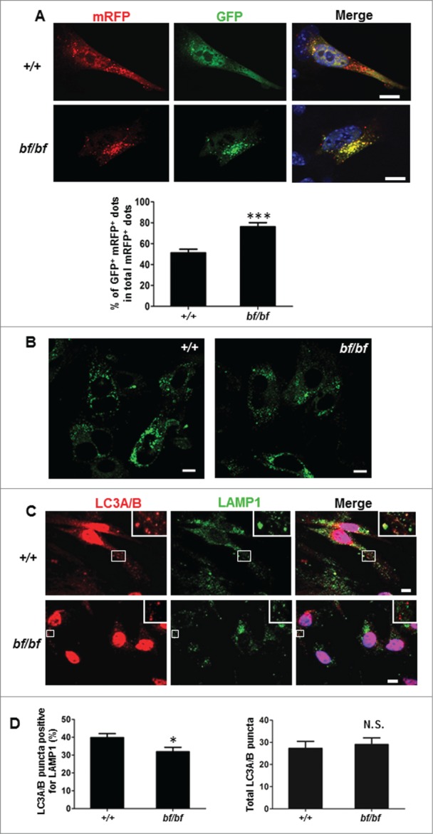
Fusion between autophagsomes and lysosomes was impaired in bf MEFs. (A) MEFs were transfected with mRFP-GFP-LC3B. After 24 h, cells were starved in DMEM without amino acids for 2 h before fixation and analyzed by confocal microscopy. Quantification data are presented as the percent of yellow puncta (autophagosomes) per total red puncta (autophagosomes and autolysosomes) from 20 randomly selected images. DAPI staining of nuclei is labeled in blue. Scale bar: 10 μm. ***P< 0.0001. (B) MEFs were stained with LysoSensor™ Green DND-189 for 30 min to observe the acidification of lysosomes. There was no apparent change in lysosomal acidification in bf MEFs compared with wild-type cells. Scale bar: 10 μm. (C) The colocalization of endogenous autophagosomal marker LC3A/B and lysosomal marker LAMP1 was reduced in bf MEFs. Cells were incubated with lysosomal inhibitors (E-64, pepstatin A and leupeptin) for 24 h and starved for additional 2 h before fixation. (D) The percentage of LC3A/B puncta colocalizing with LAMP1 relative to total LC3A/B puncta was lower in bf MEFs from 30 cells (left panel). *P < 0.05 (P = 0.0241). The total counted LC3A/B puncta were not significantly changed in bf MEFs compared with the wild-type MEFs. N.S., not significant (P = 0.7052). Scale bar: 10 μm.
