Figure 2.
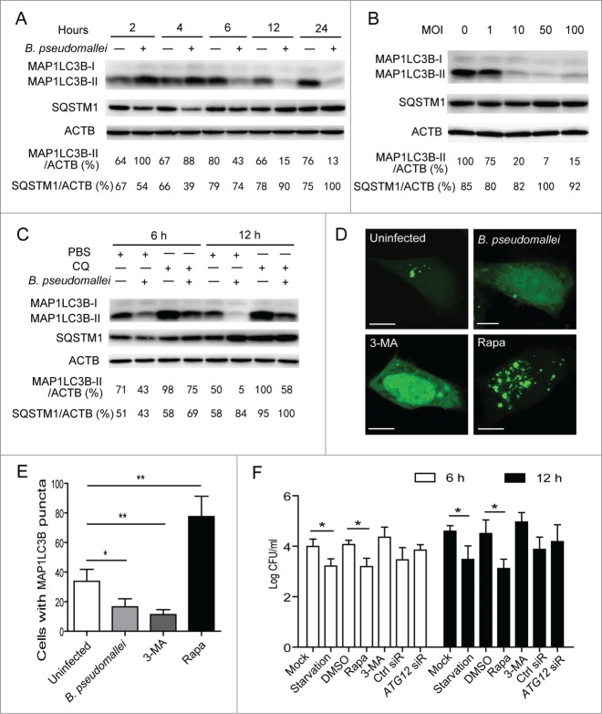
Autophagy is inhibited in response to B. pseudomallei infection. (A and B) B. pseudomallei decreased the conversion of MAP1LC3B-I to MAP1LC3B-II in A549 cells. A549 cells were treated with B. pseudomallei (MOI = 10:1) for 2, 4, 6, 12 and 24 h, or at MOI = 0, 1, 10, 50, and 100 for 6 h. (C) A549 cells were treated with B. pseudomallei (MOI = 10:1) for 6 h and 12 h in the presence of CQ (10 μM). (D and E) Confocal images show GFP-MAP1LC3B distribution in A549 cells infected with B. pseudomallei for 6 h, and uninfected cells treated with 3-MA (10 mM) or Rapa (200 nM). Scale bars: 5 μm. The number of GFP-MAP1LC3B puncta in each cell was counted. (F) Stimulation of autophagy suppresses intracellular survival of B. pseudomallei in A549 cells. After pretreatment by DMSO, Rapa and 3-MA, or transfected with ATG12 siRNA (100 nM), A549 cells were infected with B. pseudomallei (MOI = 10:1) for 6 h and 12 h. The bar represents the mean ±SD of 3 experiments. *, P<0.05, **, P<0.01.

