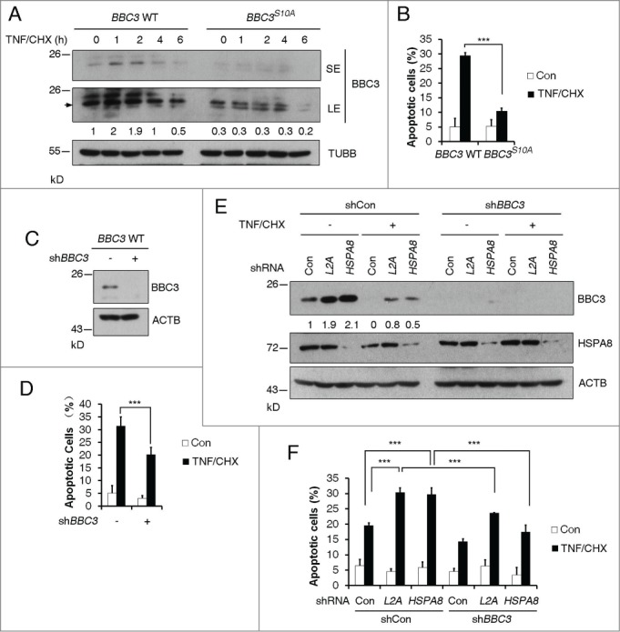
Figure 5.

BBC3 stabilization resulting from Ser10 phosphorylation or CMA blockage is essential for TNF-triggered apoptosis. (A) Representative western blots (n=3) of BBC3 WT and BBC3S10A HCT116 cells cotreated with 10 ng/ml TNF and 2.5 μg/ml CHX for the indicated time periods. BBC3 is indicated with an arrow. SE, short exposure; LE, long exposure. Quantification of BBC3 protein levels was done relative to TUBB. (B) FACS analysis data detecting early apoptotic cells showing that BBC3S10A cells confer resistance to TNF-triggered apoptosis under TNF and CHX cotreatment for 7 h. (C) Western blots showing shRNA knockdown efficiency of BBC3 in BBC3 WT HCT116 cells. (D) FACS analysis of early apoptotic cells in (C) showing that BBC3 is required for TNF-induced cytotoxicity under TNF and CHX cotreatment. BBC3 WT cells were infected with vectors containing control or BBC3 shRNA first, and 48 h after infection, cells were cotreated with TNF and CHX for 7 h before being harvested for FACS analysis. (E) Representative immunoblots (n = 3) of BBC3 WT HCT116 cells cotreated with TNF and CHX following infection with vectors containing the indicated shRNAs. BBC3 WT HCT116 cells first infected with vectors containing shCon or BBC3 shRNAs, then with shCon, LAMP2A or HSPA8 shRNAs were cotreated with TNF and CHX for 5 h and then harvested. The BBC3 protein levels were quantified relative to ACTB. (F) FACS analysis of early apoptotic cells in response to the treatment shown in (E) showing that CMA blockage could sensitize TNF-induced apoptosis,which is protected by BBC3 depletion. Statistics are depicted as mean ± SEM; P < 0.001***, n = 4, t test.
