
Figure 3.
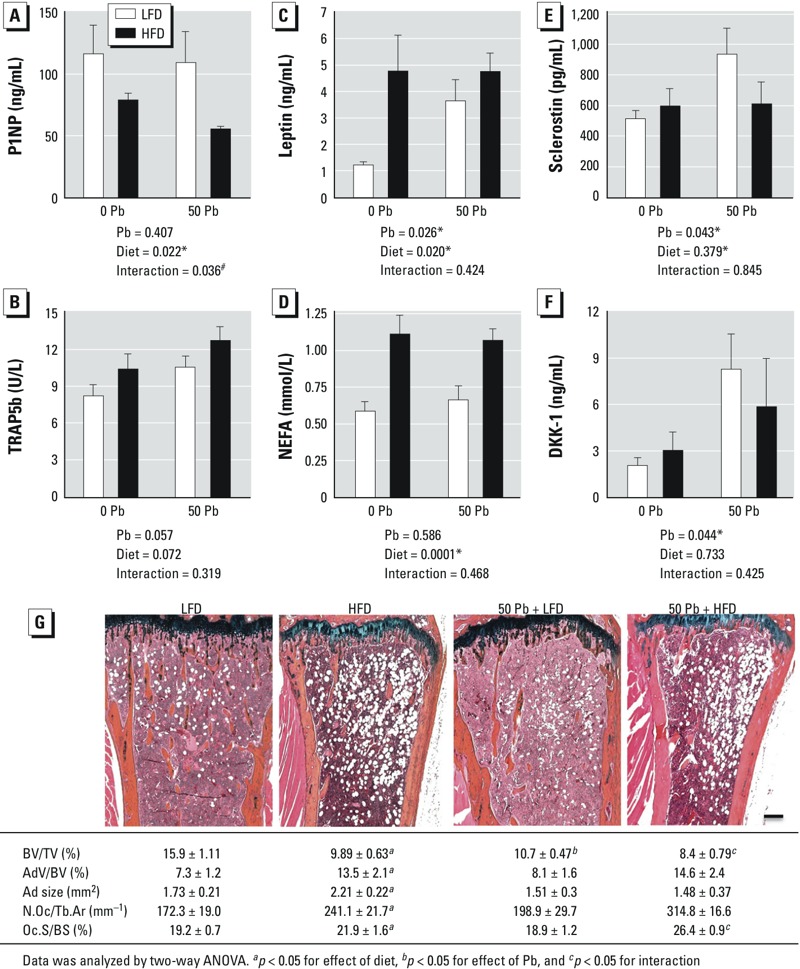
Effects of HFD and Pb (50 ppm) on histologic and serum bone parameters; Pb-exposed and control mice were placed on LFD or HFD for 6 weeks. Serum bone formation marker P1NP (A), resorption marker TRAP5b (B), leptin (C), NEFA (D), sclerostin (E), and DKK1 (F) were measured using ELISA methods. Data are mean ± SEM of 5 mice/group. (G) Trabecular bone in the proximal tibia was assessed histologically by Alcian blue hematoxylin/orange G staining (bar = 100 μm); images are representative sections from treatment groups, selected for approximation to the median BV/TV of its group. Abbreviations: AdV/BV, adipocyte volume/bone volume; Ad size, adipocyte size; BV/TV, bone volume/ total tissue volume; N.Oc/Tb.Ar, number of osteoclasts/trabecular bone area; Oc.S/BS, osteoclast surface/bone surface. The effects of Pb and/or HFD on osteoclast and adipogenic parameters were calculated and are presented at the bottom of each image. Data represent mean ± SEM for 3 mice/group. *p < 0.05 for effect of Pb or diet. #p < 0.05 for interaction of Pb and diet.