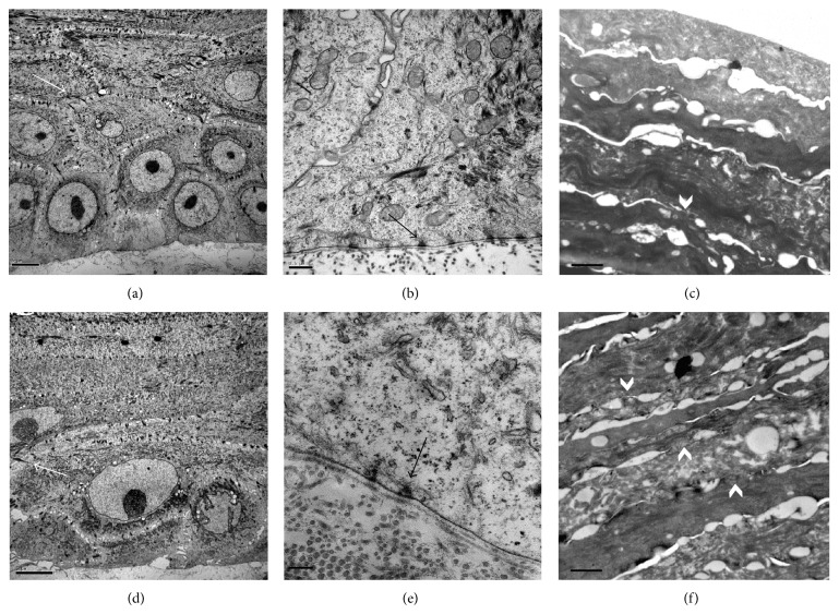
Figure 4.
Ultrastructural images of non-treated (a, b, c) and treated skin equivalent with essential oils and polyunsaturated fatty acids (d, e and f). Desmosomes between adjacent corneocytes (a, c) (white arrows), hemidesmosomes (b, e) (black arrows), and stratum corneum with vertical cohesion between corneocytes layers (white arrow head) (c, f) were observed in both samples. Scale bars: 5 μm (a and d), 0.5 μm (b), 1 μm (c and f), and 0.2 μm (e).

