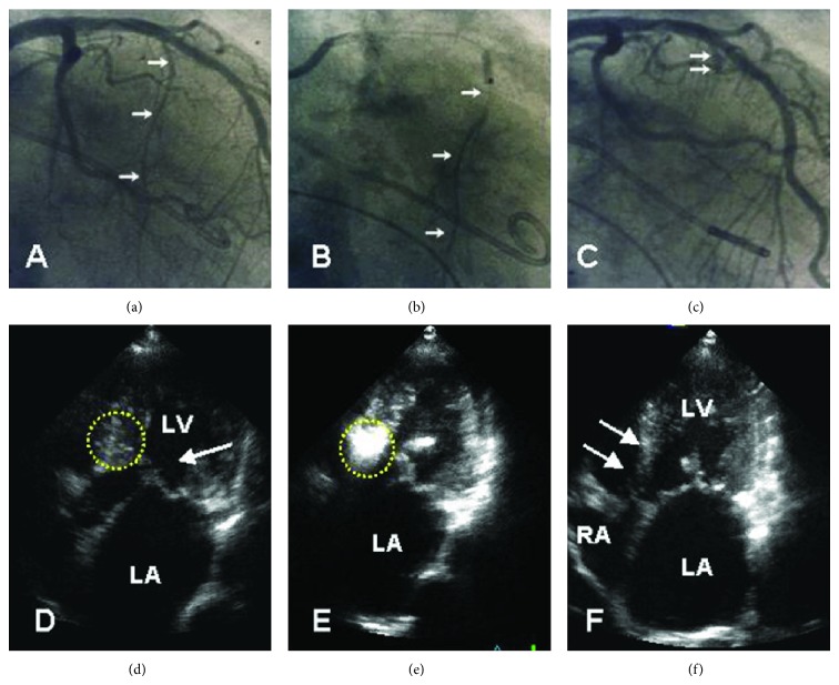
Figure 5.
Angiographic ((a)–(c)) and echocardiographic ((d)–(f)) aspect of an echotargeted septal ablation procedure (in our practice denominated as PTSMA). A guidewire is advanced into the target vessel (arrows in (a)). Subsequently, an over-the-wire balloon is introduced. The correct position and fit of the balloon are verified by contrast injection (arrows in (b)) through the central catheter lumen. The vessel stump after alcohol injection and removal of the balloon is shown in (c) (arrows). In (d), the dotted circle marks the septal target area including the SAM-septal contact zone. Contrast injection into the target vessel (e) precisely highlights this area. After 3–6 months, akinesia and thinning of the subaortic septum are clearly visible, comparable to a myectomy trough.

