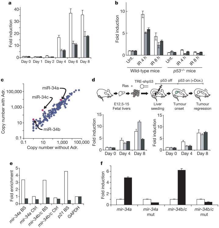
Figure 2. Genes encoding miR-34 are direct targets of p53.
a, miR-34 levels were measured in MEFs expressing a tetracycline-repressible p53 shRNA6 at the indicated times after the addition of doxycycline. White columns, mature miR-34a; grey columns, mature miR-34b; black columns, mature miR-34c. b, Wild-type and p53−/− animals were subjected to 6 Gy of ionizing radiation (IR), and miR-34 levels (identified as in a) were measured in spleens by Taqman assays both before and at the indicated times after irradiation. Unt., unirradiated. c, A group of 191 miRNAs and selected miRNA* sequences were quantified by QRT-PCR in TOV21G cells before and after treatment with 0.1 μg ml−1 adriamycin (Adr.). Results are presented in a logarithmic-scale dot plot of copy number per cell. The full data set is presented in Supplementary Table S1. d, Hepatocellular carcinomas were produced by combined expression of activated Ras and a conditional p53 shRNA13. p53 suppression was relieved by treatment with doxycycline (Dox.). Tumours were harvested at the indicated times during treatment with doxycycline, and levels of mature miR-34 were measured by Taqman assays. Levels are plotted with respect to tumours before p53 reactivation. Left: white columns, pri-mir-34a; grey columns, pri-mir-34b/34c; black columns, mp21. Right: column colours as in a. e, ChIPs were performed with p53 antibodies on wild-type MEFs (white columns) or p53−/− MEFs (black columns) treated with adriamycin. BS indicates quantification of the fragment containing the predicted p53 binding site in the mir-34a, mir-34b/c or p21 promoter regions, and Ctrl indicates a 3′ fragment from the same gene. Signals were normalized to glyceraldehyde-3-phosphate dehydrogenase (GAPDH) for each genotype. f, Firefly luciferase coding sequences were placed under the transcriptional control of human mir-34a or mir-34b/c promoter elements containing either wild-type or mutant (as indicated) p53 binding sites. These reporters were co-transfected with either control (white columns) or human p53 expression plasmids (black columns). Transfections were normalized by using a simultaneously delivered Renilla luciferase expression plasmid, pRLTK. In all cases, error bars indicate s.d. (n = 3).

