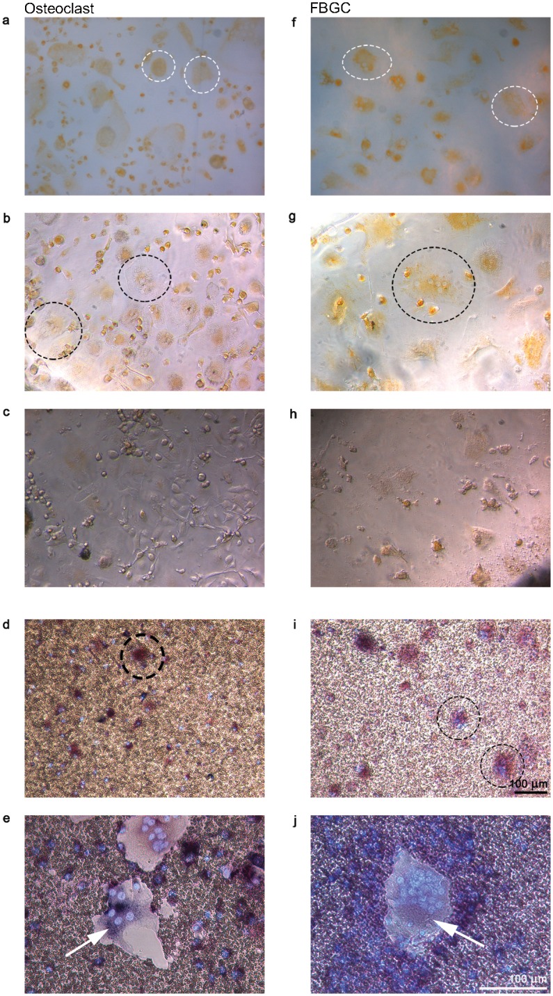
Fig 7. Resorption activity of osteoclasts and FBGCs on biomimetic hydroxyapatite coatings after concanamycin A treatment.

Cells were cultured for 25 days and stained with acridine orange to visualize sites with low pH. Osteoclasts (left column) and FBGCs (right column) stained positive for acridine orange on both bone (a, f; white dashed circles) and tissue culture plastic (b, g; black dashed circles). After incubation with concanamycin A, acridine orange-positive vacuoles were hardly detected (c, h); moreover, dissolution of hydroxyapatite was blocked (d, I; osteoclasts and FBGCs are visible in black dashed circles) compared to control cells cultured without concamycin A (e, j). Cells were stained for TRAcP and DAPI. Scale bar = 100 μm.
