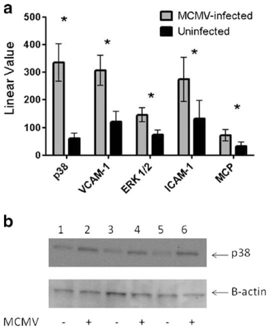
Fig. 2.
mRNA transcripts in aortas of MCMV-infected vs. uninfected Apo E KO mice (value is average of n=10 mice) at 2.5 months post-infection. a Real-time PCR was performed on cDNA using primers/fluorogenic probes specific for each gene and analysis done as described in “Materials and Methods” section; b validation of phosphorylated p38 expression by Western blot at 2.5 months post-infection: lanes 1, 3, and 5 protein patterns from aortas of uninfected Apo E KO mice; lanes 2, 4, and 6 protein patterns from aortas of MCMV-infected Apo E KO mice. Figure is representative of n=5 MCMV-infected and five uninfected control Apo E KO mice. Average levels of phosphorylated p38 protein were ~1.7-fold higher in aortas from MCMV-infected vs. uninfected Apo E KO mice. Significance (*) at p≤0.05

