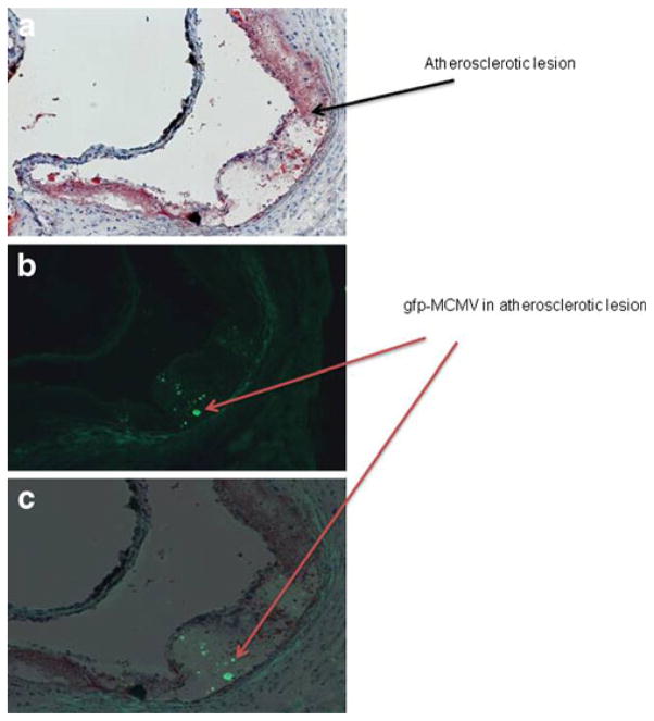
Fig. 4.
Atherosclerotic lesions in aortas of gfp-MCMV-infected Apo E KO mice at 2.5 months post-infection. a Lipid ORO-stained section; b consecutive section under fluorescence microscopy; c section B superimposed over section A, showing localization of GFP staining in the atherosclerotic plaque. Magnification ×20

